Novel Computational Phase Imaging Techniques
Phase imaging, or more precisely, imaging relative differences in optical path length, is an important tool with many applications. For example, many biological specimens of interest are nearly transparent, and phase imaging is one method by which such specimens can be imaged noninvasively with high contrast. Recent developments in optimization as well as ever-increasing computational power enable novel phase imaging techniques based on solving inverse problems modeled directly after first principles. For example, the factored form descent solver from coherence retrieval can be extended to explicitly enforce coherence and thus can be used for phase retrieval from measurements through arbitrary (known) optical systems.
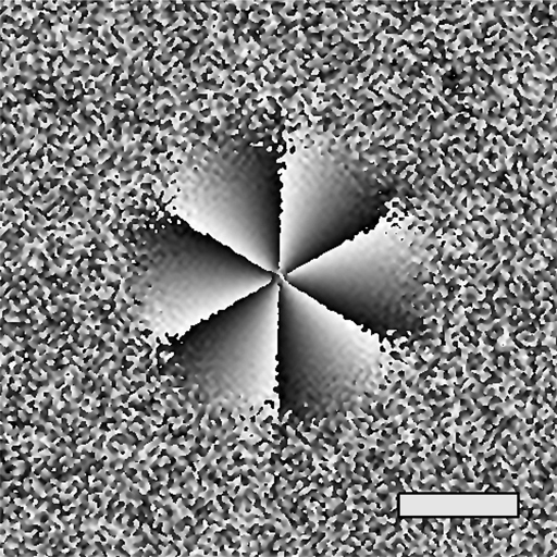
(a)
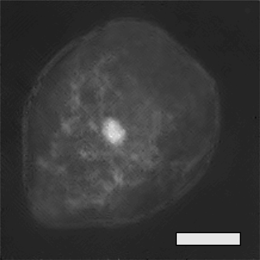
(b)

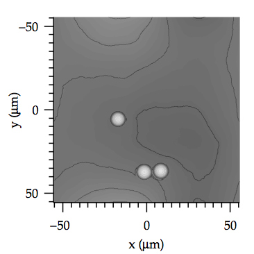
(c)
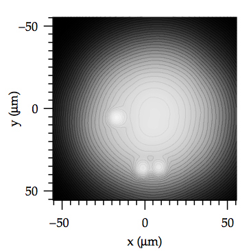
(d)
(e)
Relevant Articles and Conference Proceedings
- Zhengyun Zhang, Wensheng Chen, and George Barbastathis, "Phase Imaging Using a Hybrid Approach: Combining Wavefront Sensing With the Transport-of-Intensity Equation," in Digital Holography and Three-Dimensional Imaging, OSA Technical Digest (online) (Optical Society of America, 2015), paper DT1A.5
- Zhengyun Zhang, Zhi Chen, Shakil Rehman, and George Barbastathis, "Phase imaging using shifted wavefront sensor images," Opt. Lett. 39, 6177–6180 (2014)